Home
/ Cross Section Of A Compact Bone : Compact Bone Black And White Stock Photos Images Alamy - Compact bone (cross section of dried bone).
Cross Section Of A Compact Bone : Compact Bone Black And White Stock Photos Images Alamy - Compact bone (cross section of dried bone).
Cross Section Of A Compact Bone : Compact Bone Black And White Stock Photos Images Alamy - Compact bone (cross section of dried bone).. Hard, dense outer region • nutrient artery passes through compact bone to supply internal structures of long bone. Observe that the matrix of the bone is deposited in concentric layers that are called lamellae (5). Canaliculi allow the passage of interstitial fluid between the central canal and the lacunae housing osteocytes. Hi all, i have uploaded a new medical animation tutorial. The spongy and compact bone tissue in the cross section of a skull bone.
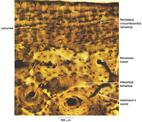
This model shows a cross section of compact bone. (b) in this micrograph of the microscopic structural unit of compact bone is called an osteon , or haversian system. Dry bone is cut and polished before mounting on a slide. Concentric layers of bone cells (osteocytes) and bone matrix surround. In the center of each osteon is the central canal, a space that houses blood vessels and nerves that supply bone.
Cartilage And Bone Basic Anatomy And Physiology Teaching Science Firefighter Paramedic from i.pinimg.com Is this possible, and if so what are the effects? A diagrammatic view of a cross section of bone. Observe that the matrix of the bone is deposited in concentric layers that are called lamellae (5). This model shows a cross section of compact bone. • structure of a long bone (continued): The outlined area is a cross section of an osteon of compact bone. Canaliculi allow the passage of interstitial fluid between the central canal and the lacunae housing osteocytes. Spongy bone and compact bone.
Hi all, i have uploaded a new medical animation tutorial.
Is this possible, and if so what are the effects? Select different colors for the. In the center of each osteon is the central canal, a space that houses blood vessels and nerves that supply bone. Cross section of the compact bone. Bone must be decalcified (by exposure to strong acids) so it can be cut into thin sections. Compact bones make up 80 percent of the human skeleton; To support the whole body, protect organs, provide. The remaining material is mostly collagen. Each osteon is composed of concentric rings of calcified. Cross section of compact bone. Canaliculi allow the passage of interstitial fluid between the central canal and the lacunae housing osteocytes. A cross section of a compact bone shows concentric circles called lamellae. This is a cross section through decalcified bone.
Bone decalcification is the removal of the mineral component using an acid, leaving the bone soft and easy to cut. In three dimensions an osteon is cylindrical in shape. Spongy bone is the osseous tissue, which fills the interior cavity of bones, consisting of mineralized bars called trabeculae. Cross section of compact bone. Compact bones make up 80 percent of the human skeleton;
Anatomy Atlases Atlas Of Microscopic Anatomy Section 1 Cells from www.anatomyatlases.org Compact bone is very different from the other tissues you have seen. A uniform cross section is the cross section of the solid, parallel to base, such that the resulting figure has the same shape and size as that of the base of the figure.more about uniform cross sectionsolids like. Concentric layers of bone cells (osteocytes) and bone matrix surround. There are trabeculae in spongy bone which gives its sponge like appearance. They build the entire picture, improve your understanding, consolidate the information and facilitate recall. Observe that the matrix of the bone is deposited in concentric layers that are called lamellae (5). The spongy and compact bone tissue in the cross section of a skull bone. Compact bone, also known as cortical bone, is a denser material used to create much of the hard structure of the skeleton.
Each osteon is composed of concentric rings of calcified.
Select different colors for the. Observe that the matrix of the bone is deposited in concentric layers that are called lamellae (5). Compact bone is very different from the other tissues you have seen. A diagrammatic view of a cross section of bone. The spongy and compact bone tissue in the cross section of a skull bone. Cross section of compact bone. A cross section of a compact bone shows concentric circles called lamellae. The outlined area is a cross section of an osteon of compact bone. This model shows a cross section of compact bone. Compact bones make up 80 percent of the human skeleton; They build the entire picture, improve your understanding, consolidate the information and facilitate recall. To support the whole body, protect organs, provide. Bone decalcification is the removal of the mineral component using an acid, leaving the bone soft and easy to cut.
To support the whole body, protect organs, provide. Is this possible, and if so what are the effects? As the names suggest compact bone looks compact and the spongy bone looks like sponges. Compact bone, makes up the dense material in a long section of a bone. The spongy and compact bone tissue in the cross section of a skull bone.
Plos One Spectroscopic Studies On Organic Matter From Triassic Reptile Bones Upper Silesia Poland from journals.plos.org A diagrammatic view of a cross section of bone. Cortical or compact bone can be distinguished macroscopically from cancellous or trabecular bone. The two layers of compact bone and the interior spongy bone work together to protect the internal organs. Canaliculi allow the passage of interstitial fluid between the central canal and the lacunae housing osteocytes. This is a cross section through decalcified bone. The remaining material is mostly collagen. Compact bone (cross section of dried bone). Observe that the matrix of the bone is deposited in concentric layers that are called lamellae (5).
Cortical or compact bone can be distinguished macroscopically from cancellous or trabecular bone.
There are two ways to study bone histology. Concentric layers of bone cells (osteocytes) and bone matrix surround. Sclerostin inhibits bone formation mostly by antagonizing lrp5/6, thus inhibiting wnt signaling. This is a cross section through decalcified bone. And the the femur is the thigh bone, the longest bone in the body. Compact bone, also known as cortical bone, is a denser material used to create much of the hard structure of the skeleton. Compact bone is very different from the other tissues you have seen. Hi all, i have uploaded a new medical animation tutorial. For example, in the human cancellous bone in which the cross struts are disconnected (c) is not very capable of supporting a. Bone decalcification is the removal of the mineral component using an acid, leaving the bone soft and easy to cut. (b) in this micrograph of the microscopic structural unit of compact bone is called an osteon , or haversian system. There are trabeculae in spongy bone which gives its sponge like appearance. Cross section of compact bone.
Osteocyte processes lie in tiny canals (canaliculi) in the bone matrix cross section of a bone. A cross section of a compact bone shows concentric circles called lamellae.

.)